|
Cycle 1 (2011 Deadline)
Characterization of cassava mosaic gemini viruses and their satellites in cassava at the cellular level PI: Joseph Ndunguru, Mikocheni Agricultural Research Institute US Partner: Linda Hanley-Bowdoin, North Carolina State University Project Dates: May 2012 - August 2014 Project Overview Cassava is an important staple crop in Africa and Asia, where it is eaten by more than 700 million people every day. It is grown by subsistence farmers in the poorest villages and is often the only food source when other crops fail or are destroyed by conflict. Cassava can grow under drought, high temperature, and poor soil conditions, but its production is severely limited by viral diseases. Cassava mosaic disease (CMD) is caused by a DNA virus complex consisting of seven geminivirus species that act synergistically to enhance disease severity. Recently, two satellite DNAs associated with the complex have been shown to break resistance and enhance symptoms. 
| 
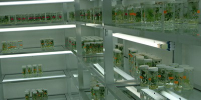
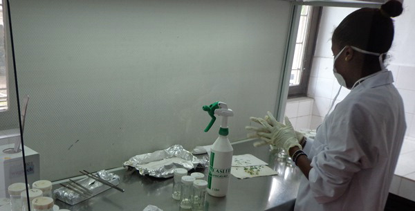
| | Cassava plants maintained in a tissue culture lab ready for sub-culturing at MARI. (Photo courtesy Joseph Ndunguru). | A research student getting prepared for sub-culturing of cassava plants at MARI(Photo courtesy Joseph Ndunguru). |
Cassava mosaic geminiviruses (CMGs) induce diverse symptoms in cassava depending on the host genotype, age of infection, amount of virus inoculum, virus strain, vector activity, and environmental and other host factors. Research on CMGs has generated extensive information about viral diversity, genome sequence, replication, transmission, disease epidemiology and disease control. In contrast, there are no reports that describe the changes that occur in cassava leaves at a cellular level in response to CMG infection. The goal of this proposal is to establish cell biology infrastructure and expertise at Mikocheni Agricultural Research Institute (MARI) in Tanzania and to use these resources to characterize CMG infection in cassava. A combination of light and fluorescent microcopy will be used to examine CMD processes at a cellular level in cassava leaves. Using in situ hybridization to detect CMG and satellite DNAs in cassava leaves, these researchers will seek to determine if different CMGs infect different leaf cell types and if the nature and number of the target cells change in mixed infections and/or in the presence of the satellites. The research team will also examine the cellular architecture of infected leaves as a first step toward understanding the physiological basis of the extreme leaf deformation phenotypes correlated with the presence of CMD-associated satellites. The application of cell biology to CMD represents a unique opportunity to study the interactions of different viruses with a common host and with each other and satellite DNAs. The increased knowledge to be gained through the project should contribute to understanding of this important plant virus and to the development of sustainable strategies to control it and thereby limit its economic and nutritional impact. Overall Summary of Activities
This project proved to be successful both locally and nationally. The information generated from this project has helped to design cassava virus disease diagnostic tools resulting in efficient and accurate disease diagnostics to inform the formulation of disease management strategies. Proper virus diagnostics have resulted in the development of virus distribution maps in the country that are now used by decision makers to decide: a) where to deploy clean cassava planting materials; b) where to evaluate cassava materials (hot spot areas); c) where to multiply clear cassava materials (low disease pressure areas); and d) how to strengthen plant health regulations by restricting unsafe movement of cassava germplasm. Furthermore, cassava farmers in Tanzania have received virus-free planting materials that have resulted into a significant yield increase and the microscopy has enabled regular virus monitoring in the planting material.
Locally, both the human and infrastructural capacity at MARI was significantly enhanced through training of local scientists and acquisition of the lab equipment. University students who should have gone abroad to conduct their research work are now conducting their research at MARI. With this, more students can be trained and the availability of the microscopy facility at MARI has inspired many young scientists to undertake research in the areas of biotechnology, including disease diagnostics using molecular techniques, as well as detection of plant viruses using the immuno and insitu hybridization techniques. Finally, this project also has stimulated more collaboration among MARI research scientists and other institutions within and outside Tanzania.

| 
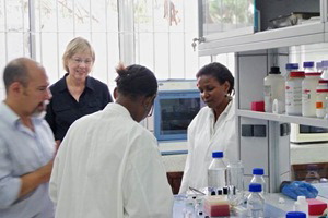
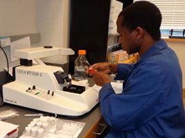
| | Scientists from Mikocheni Agricultural Research Institute (MARI) are being trained on to use the vibratome and fluorescent microscope (Photo courtesy Joseph Ndunguru). | Linus Paul, who is in charge of microscopy at MARI, is working with a vibratome (Photo courtesy Joseph Ndunguru). |
Back to PEER Cycle 1 Grant Recipients
| 


